
A slow response to persistent force
Epithelial tissues line surfaces and cavities throughout the body, where they must accommodate repeated deformation without losing their integrity. Their cells contain keratin intermediate filaments: a mechanically robust part of the cytoskeleton that helps distribute forces within and between cells. Under ordinary conditions, keratin forms a network at the cell edge and around the nucleus, with filament connections linking these regions.
A study published on 27 July in Nature Physics reports that this arrangement can be dramatically remodelled under large, sustained stretching. Rather than responding instantly, keratin in epithelial monolayers slowly coalesced into thick, star-shaped bundles. The transition culminated in what the researchers call nuclear uncaging: the nucleus moved out of the three-dimensional keratin network that initially surrounded it.
The result adds a time-dependent dimension to the mechanics of epithelia. Keratin is often described as a protective scaffold, but the work suggests that its response to force is not simply to remain in place and resist deformation. At sufficiently high and prolonged strain, the network can reorganise across many cells and change how force is coupled to the nucleus.
From cellular rims to supracellular cables
The researchers studied monolayers of MDCK cells, a widely used epithelial cell line derived from canine kidney, in two complementary settings. First, they examined pressure-driven domes that arise when fluid accumulates beneath a cell layer. They also used a device that applied constant pressure to curved epithelial monolayers while allowing hours of live imaging.
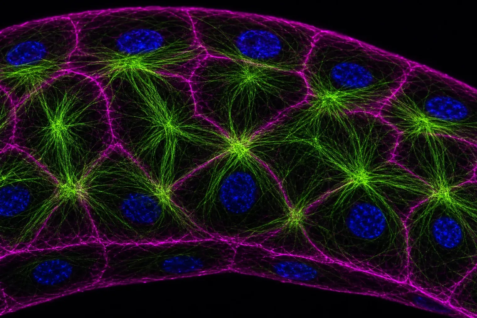
Before deformation, keratin displayed the expected rim-and-spoke architecture: a peripheral network associated with the cell cortex, linked by shorter bundles to a mesh surrounding the nucleus. Under persistent stretching, however, keratin first became depleted near tricellular junctions, where three cells meet. The displaced material then accumulated in thicker bundles crossing cell-cell junctions along the opposite direction.
Individual cells thereby acquired a star-like pattern, with long bundles radiating from a dense central region. Importantly, matching bundles connected across neighbouring cells. What began in scattered cells expanded into multicellular clusters, eventually forming a percolated keratin network at the tissue scale.
This is a significant distinction. A cell’s cytoskeleton is often considered primarily at the single-cell level, but epithelial keratin networks are connected through intercellular junctions. The observed transition therefore links subcellular filament rearrangement to a collective, supracellular architecture capable of redistributing mechanical loads across a monolayer.
Why tension can lead to compression
The most counterintuitive finding concerns the nucleus. One might expect tension applied to a tissue to pull directly on the nucleus. Instead, the team’s computational model indicated that increasing tension in the entangled keratin network generates a growing steric, or contact-based, compressive pressure around it.
As keratin fibres reorganised into radial bundles, they progressively lost their coverage over the top of the nucleus and concentrated below it. The simulations predicted that the tightening network would press on the nucleus before a rapid escape from the keratin enclosure relieved that pressure. Live confocal imaging supported this sequence: the fluorescent signals from DNA and keratin, initially aligned in depth, increasingly separated as the keratin reorganised and the nucleus shifted relative to it.
The term “escape” should not be read as implying that a nucleus moves freely through the cell. It describes a geometrical and topological change in which the nucleus is no longer enveloped by the keratin mesh. In the final star-bundled configuration, the dense keratin knot was positioned on the basal side of the cell, while the nucleus lay above it and retained only limited surrounding keratin.
The authors describe the mechanism as compression by tension. It is an instructive example of how force transmission in complex cellular materials can differ from simple intuition: pulling on the outer tissue can, through the architecture of an internal network, squeeze a structure inside the cell.
Actin sets the pace
The remodelling was notably slow. In the pressure-controlled experiments, the average time to meet the study’s criteria for bundling was roughly seven hours, although cells varied substantially. The star-like arrangement emerged only after an earlier stage in which keratin-depleted regions developed near tricellular junctions.
Experiments disrupting the link between actin and keratin accelerated the process. The researchers targeted plectin, a crosslinking protein that helps connect keratin to the actin cortex and, indirectly, to the nuclear surface. Cells expressing a dominant-negative plectin construct underwent the same broad transition but much faster than control cells. Disrupting actin with a drug also prompted rapid keratin bundling, although not the complete untreated star-shaped organisation.
These tests suggest that interactions with the actin cortex restrain or regulate keratin’s reorganisation under sustained strain. They do not mean that actin is dispensable from epithelial mechanics; rather, the findings identify the actin-keratin linkage as a key controller of the timescale on which this specific keratin transition occurs.
Protection may involve changing the route of force
Previous research has shown that keratin networks can protect epithelial monolayers at large deformation by contributing to strain stiffening, and that keratin-linked structures can shield nuclei from mechanical deformation. The new study complicates that picture by revealing a condition in which the perinuclear keratin arrangement is abandoned.
Whether uncaging leaves the nucleus more vulnerable or provides an alternative form of protection remains unresolved. A nucleus without a close keratin cage could be exposed more directly to deformation. Conversely, separating it from a highly stressed cytoskeletal network might reduce transmission of damaging forces. The study did not measure DNA damage, nuclear-envelope rupture, gene-expression changes or longer-term cell fate after uncaging, so those possibilities remain hypotheses for future experiments.
There are also limits to how far the result can be generalised. The evidence comes from engineered and cultured MDCK epithelial monolayers exposed to substantial, prolonged deformation. Different epithelia, extracellular matrices, developmental settings and disease states may alter the threshold, timing or outcome of the response.
Even with those caveats, the work provides a detailed physical framework for studying how tissues adapt when force persists rather than arrives as a brief pulse. It shows that keratin can function not only as a passive reinforcement system but also as a dynamic, collective structure whose rearrangement changes the mechanical environment of the nucleus itself.
Sources
- Dynamics of supracellular keratin bundling and nuclear uncaging in stretched epithelia — Nature Physics
- Under pressure: when tension builds up, the nucleus escapes — Institute for Bioengineering of Catalonia
- Rupture strength of living cell monolayers — Nature Materials
- The laminin–keratin link shields the nucleus from mechanical deformation and signalling — Nature Materials


